OCT Scanning - Axial resolution: 5 μm (in tissue)
- Transverse resolution: 15 μm (in tissue)
- Scan speed: 27,000 A-scans per second
- A-scan depth: 2.0 mm (in tissue), 1024 points
- Optical source: superluminescent diode (SLD), 840 nm
Fundus Imaging - Line scanning ophthalmoscope (LSO)
- Live during scanning
- Transverse resolution: 25 μm (in tissue)
- Optical source: superluminescent diode (SLD), 750 nm
- Field of view: 36° x 30°
Scan Patterns - Macular Cube 200 x 200 Combo: 200 horizontal scan lines comprised
of 200 A-scans
- Macular Cube 512 x 128 Combo: 128 horizontal scan lines comprised
of 512 A-scans
- 5 Line Raster: 4096 A-scans per B-Scan (adjustable length, spacing and
orientation)
Focus Adjustment Range - –20D to +20D (diopters)
Fixation - Internal and external
Computer - Windows® XP Pro
- High-performance multi-core processor
- Internal storage: > 80,000 scans
- CD-RW, DVD-ROM drive
- Integrated 15” color flat panel display
Pupil Size Requirement - ≤ 2.0 mm (≥ 3.0 mm optimal for LSO)
Dimensions/Weight
(Instrument Only)
- 25.6 L x 17.3 W x 20.9 H (in); 65 L x 44 W x 53 H (cm)
- 83 lbs; 37.6 kg
Electrical 100–120V~, 50/60Hz, 5A
220–240V~, 50/60Hz, 2.5A
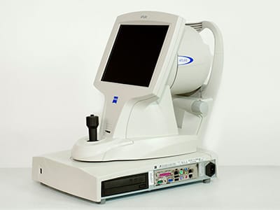
Carl Zeiss Humphrey Atlas 995 is the most innovative tool topographer come with advanced imaging and analysis technology software Humphrey Atlas 995 come with the computer linked to a lighted bowl so patients can sit in front of the bowl During a diagnostic test. RME: Downloads.
- The Zeiss Atlas 995 Corneal Topographer is the most advanced system available. The Zeiss Atlas 995 offers ultra-low illumination and increased peripheral coverage. The Zeiss Atlas 995 is ideal for high volume corneal and contact lens specialists who require comprehensive and detailed peripheral corneal and pupil assessments. The greatest advantage of corneal topography is its.
- The Zeiss Atlas 995 Corneal Topographer is the most advanced system available. The Zeiss Atlas 995 offers ultra-low illumination and increased peripheral coverage. The Zeiss Atlas 995 is ideal for high volume corneal and contact lens specialists who require comprehensive and detailed peripheral corneal and pupil assessments.
- View & download of more than 391 Zeiss PDF user manuals, service manuals, operating guides. Microscope, Binoculars user manuals, operating guides & specifications.
Carl Zeiss Meditec is an international giant in the field of manufacturing and distributing high-end optics and optical products.
The greatest advantage of corneal topography is its ability to detect irregular conditions of your cornea invisible to most conventional testing. And because the computer can save your exam information, Dr. Seibel can monitor any changes to your cornea and/or your corneal stability over time.


Corneal topography is a computer assisted diagnostic tool that creates a three-dimensional map of the surface curvature of your cornea. The Zeiss Atlas 995 Topographer is the most innovative tool of its kind.
How does it work?
The cornea (the front window of the eye) is responsible for about 70 percent of the eye’s focusing power. An eye with normal vision has an evenly rounded cornea, but if the cornea is too flat, too steep, or unevenly curved, less than perfect vision results.

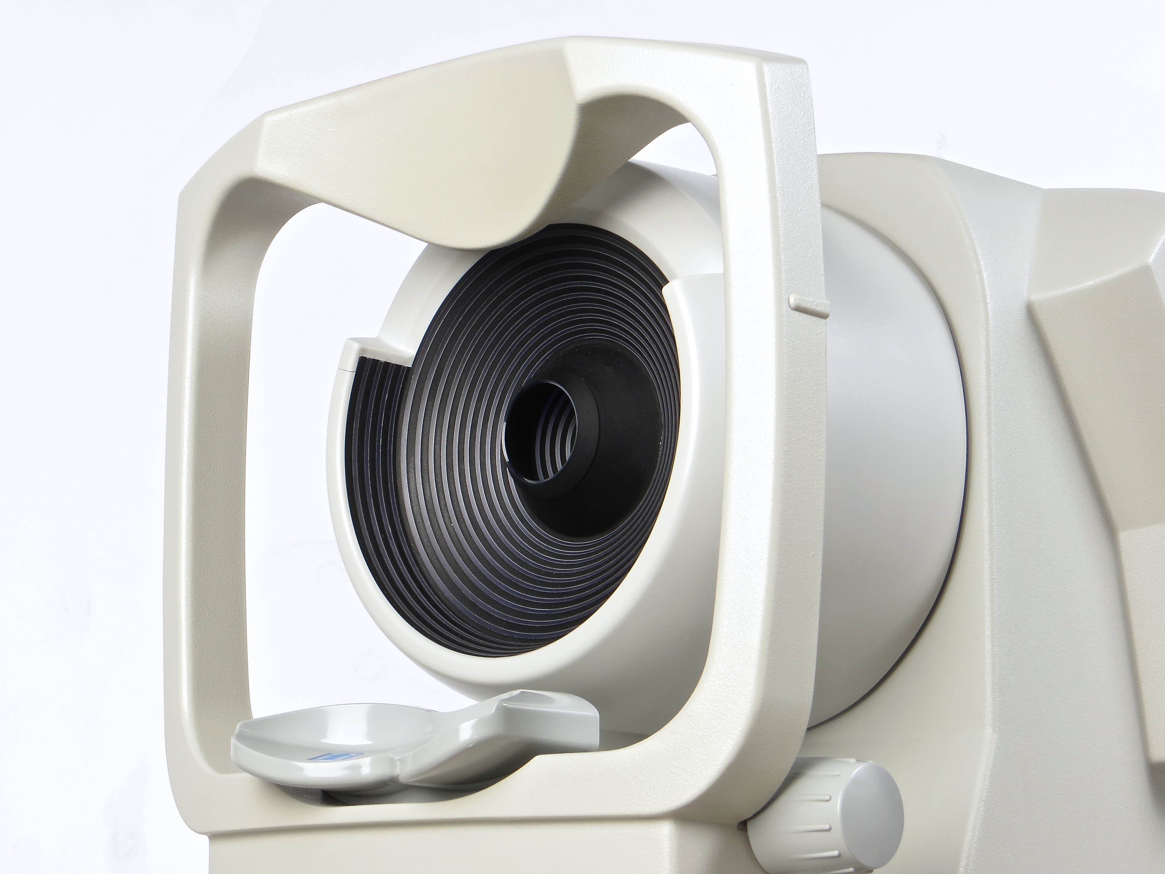
The Atlas 995 corneal topographer consists of a computer linked to a lighted bowl that contains a pattern of rings. During a diagnostic test, you would sit in front of the bowl with your head pressed against a bar while a series of data points are generated. Computer software digitizes these data points to produce a printout of the corneal shape, using different colors to identify different elevations, much like a topographic map of the earth displays changes in the land surface. This is a painless and brief non-contact test.
Zeiss Atlas 995 Manual
What do the colors mean?
Corneal mapping reveals patterns indicating conditions that may affect the selection of the best type of vision correction or treatment for you. These patterns are color-coded according to highs and lows – red being the highest and purple being the lowest, in a continuous rainbow order. A few examples of corneal color maps are shown below.
- This is a typical round cornea with a shape that changes uniformly from the center to the edge
- This is a typical astigmatism where the cornea is not round and has two balanced but different focusing powers.
- This is a asymmetric astigmatism where the cornea is not round and the focusing powers are not balanced.
- This is a keratoconus is a condition where an area of the cornea assumes a cone like shape.
Corneal topography produces a detailed, visual description of the shape and power of your cornea. This type of analysis provides Dr. Seibel with very fine details regarding the condition of your corneal surface. These details are used to diagnose, monitor, and treat various eye conditions. They are also used in fitting contact lenses and for planning surgery, including LASIK.
Zeiss Atlas Topographer
For LASIK the corneal topography map is used in conjunction with other tests to determine exactly how much corneal tissue will be removed to correct vision and with what LASIK pattern.